Recently, Deep Learning has made rapid advances in the performance of medical image analysis challenging physicians in their traditional fields. In the pathology and radiology fields, in particular, automated procedures can help to reduce the workload of pathologists and radiologists and increase the accuracy and precision of medical image assessment, which is often considered subjective and not optimally reproducible. In addition, Deep Learning and Computer Vision demonstrate the ability/potential to extract more clinically relevant information from medical images than what is possible in current routine clinical practice by human assessors. Nevertheless, considerable development and validation work lie ahead before AI-based methods can be fully ready for integrated into medical departments.
The workshop on AI-enabled medical image analysis (AIMIA) at ECCV 2022 aims to foster discussion and presentation of ideas to tackle the challenges of whole slide image and CT/MRI/X-ray analysis/processing and identify research opportunities in the context of Digital Pathology and Radiology/COVID19.
High-quality original contributions should be targeted in several contexts such as, using self-supervised and unsupervised methods to enforce shared patterns emerging directly from data, developing strategies to leverage few (or partial) annotations, promoting interpretability in both model development and/or the results obtained, or ensuring generalizability to support medical staff in their analysis of data coming from multi-centres, multi-modalities or multi-diseases.
* All times are IDT - Israel Daylight Time (GMT+3) *
Opening session
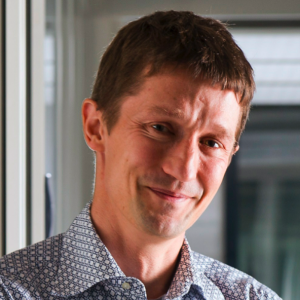
Keynote talk: Prof. Henning Müller
ExaMode, a practical approach for machine learning in digital pathology
Abstract:
The ExaMode project addresses several challenges that particularly deep learning has when using histopathology data. From creating data sets
for concrete scenarios, using weak labels as annotations and multimodal learning from the data in collaboration with hospitals, the project
really chooses a practical approach for machine learning-based decision support in digital pathology.
About Henning Müller:
Henning Müller studied medical informatics at the University of Heidelberg, Germany, then worked at Daimler-Benz research in Portland, OR, USA.
From 1998-2002 he worked on his PhD degree in computer vision at the University of Geneva, Switzerland with a research stay at Monash University,
Melbourne, Australia. Since 2002, Henning has been working for the medical informatics service at the University Hospital of Geneva. Since 2007,
he has been a full professor at the HES-SO Valais and since 2011 he is responsible for the eHealth unit of the school. Since 2014, he is also professor
at the medical faculty of the University of Geneva. In 2015/2016 he was on sabbatical at the Martinos Center, part of Harvard Medical School in Boston,
MA, USA to focus on research activities. Henning is coordinator of the ExaMode EU project and was coordinator of the Khresmoi EU project, scientific
coordinator of the VISCERAL EU project. Since early 2020 he is also a member of the Swiss National Research Council.
Coffee Break
Oral session I: Papers presentations
(see each track schedule for more details)
Keynote talk: Prof. Inti Zlobec
2022: A digital pathology odyssey
Abstract:
Some have called it the 3rd revolution in pathology: digitization and AI. On the one hand, digitization of histopathology images opens
the window of opportunity for new diagnostics tools, innovative technologies for research and bridging the gap between different medical
domains, but on the other is associated with the challenges of handling huge amounts of data, the lack of standardization and an insecurity
around the implementation of AI. How will this revolution affect personalized medicine and what is pathology of the future?
About Inti Zlobec:
Inti Zlobec is Professor of Digital Pathology (Extraordinarius) at the Institute of Pathology, University of Bern, Switzerland. Originally
from Montreal, Canada, she graduated with a PhD degree in Experimental Pathology, from McGill University before moving to Switzerland in
2007. She received her Habilitation in 2010 after a post-doctoral fellowship at the University Hospital Basel, where she conducted tissue-based
research including molecular pathology in the field of colorectal cancer using biostatistical models. Now, she leads an inter-disciplinary
research group using artificial intelligence to study pathology data and images. The aim is to discover and validate novel prognostic and predictive
biomarkers for cancer patients, and their potential implementation into diagnostic routine. Inti Zlobec is a member of the Executive Team of the
Center for Artificial Intelligence in Medicine (CAIM) of the University of Bern, Co-Founder and President of the Swiss Consortium for Digital
Pathology (SDiPath) and Chair of the European Society of Pathology (ESP) Working Group IT (Computational).
Oral session II & III: Papers presentations
(see each track schedule for more details)
Coffee Break

Keynote talk: Prof. Dimitris Metaxas
Histopathology Image Analytics for Cancer Diagnosis and Treatment using novel Deep Learning Methods
Abstract:
Histopathology plays a vital role in cancer diagnosis, prognosis, and treatment decisions. The
whole slide imaging technique that captures the entire slide as a digital image allows the
pathologists to view the slides digitally as opposed to what was traditionally viewed under a
microscope. The recent development of deep learning methods has resulted in powerful
computational methods for the quantitative and objective analyses of histopathology images,
which can reduce the intensive labor and improve the efficiency of pathologists compared with
manual examinations. In this talk we focus on deep learning based solutions for histopathology
image analysis for cancer diagnosis and treatment, specifically, nuclei segmentation for cancer
diagnosis, gene mutation and pathway activity prediction for cancer treatment. We will outline
our computational methods for annotation efficient weakly supervised and instance
segmentation nuclei segmentation algorithms with 60 times speed up. We will also present a
deep learning model to predict the genetic mutations and biological pathway activities directly
from histopathology slides in breast cancer. The weight maps of tumor tiles are visualized to
understand the decision-making process of deep learning models. Our results provide new
insights into the association between pathological image features, molecular outcomes and
targeted therapies for breast cancer patients.
About Dimitris Metaxas:
Dimitris Metaxas is a Distinguished Professor in the Computer and Information Sciences
Department at Rutgers University. He is directing the Center for Computational Biomedicine,
Imaging and Modeling (CBIM) and the NSF University-Industry Collaboration Center CARTA
with emphasis on real time and scalable data analytics, AI and machine learning methods with
applications to computational biomedicine and computer vision. Dr. Metaxas has been
conducting research towards the development of novel methods and technology upon which AI,
machine learning, computer vision, medical image analysis, and computer graphics can
advance synergistically. In medical and biological image analysis new AI, Machine Learning and
model-based methods have been developed for material modeling and shape estimation of
internal body parts (e.g., heart, lungs) from MRI, SPAMM and CT data, cancer diagnosis, cell
segmentation from histopathology images, cell tracking, cell type analysis and linking genetic
mutations to cells. Dr. Metaxas has published over 700 research articles in these areas and has
graduated 63 PhD students, who occupy prestiguous academic and industry positions. His
research has been funded by NIH, NSF, AFOSR, ARO, DARPA, HSARPA, and the ONR. Dr.
Metaxas work has received many best paper awards and he has 8 patents. He was awarded a
Fulbright Fellowship in 1986, is a recipient of an NSF Research Initiation and Career awards,
and an ONR YIP. He is a Fellow of the American Institute of Medical and Biological Engineers,
a Fellow of IEEE and a Fellow of the MICCAI Society. He has been general chair of IEEE CVPR
2014, Program Chair of ICCV 2007, General Chair of ICCV 2011, FIMH 20011 and MICCAI
2008 and the Senior Program Chair for SCA 2007.
Closing session
Opening session
Keynote talk: Prof. Henning Müller
ExaMode, a practical approach for machine learning in digital pathology
Coffee Break
Oral session I: Papers presentations
(10 min presentations + 5 min Q&A)
"CCRL: Contrastive Cell Representation Learning"
by Ramin Ebrahim Nakhli, Amirali Darbandsari, Hossein Farahani and Ali Bashashati
"Unsupervised Domain Adaptation Using Feature Disentanglement And GCNs For Medical Image Classification"
by Dwarikanath Mahapatra, Steven W Korevaar, Behzad Bozorgtabar and Ruwan Tennakoon
"FUSION: Fully Unsupervised Test-Time Stain Adaptation via Fused Normalization Statistics"
by Nilanjan Chattopadhyay, Shiv Gehlot and Nitin Singhal
"Relieving pixel-wise labeling effort for pathology image segmentation with self-training"
by Romain Mormont, Mehdi Testouri, Raphaël Marée and Pierre Geurts
"Variability Matters: Evaluating inter-rater variability in histopathology for robust cell detection"
by Cholmin Kang, Chunggi Lee, Heon Song, Minuk Ma and Sérgio Pereira
Keynote talk: Prof. Inti Zlobec
2022: A digital pathology odyssey
Coffee Break
Oral session II: Papers presentations
(10 min presentations + 5 min Q&A)
"Automatic Grading of Cervical Biopsies by Combining Full and Self-Supervision"
by Mélanie Lubrano, Tristan Lazard, Guillaume Balezo, Yaelle Bellahsen, Cécile Badoual, Sylvain Berlemont and Thomas Walter
"Using Whole Slide Image Representations from Self-Supervised Contrastive Learning for Melanoma Concordance Regression"
by Sean Grullon, Vaughn Spurrier, Corey Chivers, Yang Jiang, Jiayi Zhao, Julianna Ianni, Michael Bonham, Kiran Motaparthi and Jason Lee
"Multi-Scale Attention-Based Multiple Instance Learning for Classification of Multi-Gigapixel Histology Images"
by Made Satria Wibawa, Kwo Wai Lo, Lawrence Young and Nasir Rajpoot
"A Deep Wavelet Network for High-Resolution Microscopy Hyperspectral Image Reconstruction"
by Qian Wang and Zhao Chen
Coffee Break
Keynote talk: Prof. Dimitris Metaxas
Explainability and generalizability in medical image analysis and prediction, based on unsupersised and semi-supervised methods
Closing session
Opening session
Keynote talk: Prof. Henning Müller
ExaMode, a practical approach for machine learning in digital pathology
Coffee Break
Oral session I: Papers presentations
(10 min presentations + 3 min Q&A)
"Harmonization of Diffusion MRI Data Obtained with Multiple Head Coils Using Hybrid CNNs"
by Leon Weninger, Sandro Romanzetti, Julia Ebert, Kathrin Reetz and Dorit Merhof
"When CNN Meet with ViT: Towards Semi-Supervised Learning for Multi-Class Medical Image Semantic Segmentation"
by Ziyang Wang, Tianze Li, Jian-Qing Zheng and Baoru Huang
"Explainable Model for Localization of Spiculation in Lung Nodules"
by Mirtha L. Lucas, Miguel Lerma, Jacob Furst and Daniela Raicu
"Self-Supervised Pretraining for 2D Medical Image Segmentation"
by András Kalapos and Bálint Gyires-Tóth
"Medical Image Super Resolution by Preserving Interpretable and Disentangled Features"
by Dwarikanath Mahapatra, Behzad Bozorgtabar and Mauricio Reyes
"Medical Image Segmentation: A Review of Modern Architectures"
by Natalia E Salpea, Paraskevi Tzouveli and Dimitrios Kollias
"Multi-Label Attention Map Assisted Deep Feature Learning for Medical Image Classification"
by Dwarikanath Mahapatra and Mauricio Reyes
Oral session II: Papers presentations
(8 min presentations + 2 min Q&A)
"AI-MIA: COVID-19 Detection & Severity Analysis through Medical Imaging"
by Dimitrios Kollias, Anastasis Arsenos and Stefanos Kollias
"CMC_v2: Towards More Accurate COVID-19 Detection with Discriminative Video Priors"
by Junlin Hou, Jilan Xu, Nan Zhang, Yi Wang, Yuejie Zhang, Xiaobo Zhang and Rui Feng
"Spatial-Slice Feature Learning Using Visual Transformer and Essential Slices Selection Module for COVID-19 Detection of CT Scans in the Wild"
by Chih-Chung Hsu, Chi-Han Tsai, Guan Lin Chen, Sin-Di Ma and Shen Chieh Tai
"Using a 3D ResNet for detecting the presence and severity of COVID-19 from CT scans"
by Robert B Turnbull
"PVT-COV19D: COVID-19 Detection through Medical Image Classification Based on Pyramid Vision Transformer"
by Lilang Zheng, Jiaxuan Fang, Xiaorun Tang, Hanzhang Li, Jiaxin Fan J Fan, TIanyi Wang, Rui Zhou and Zhaoyan Yan
"CNR-IEMN-CD and CNR-IEMN-CSD Approaches for COVID-19 Detection and COVID-19 Severity Detection from 3D CT-Scan"
by Fares Bougourzi, Cosimo Distante, Fadi Dornaika and Abdelmalik Taleb-Ahmed
Coffee Break
Oral session III: Papers presentations
(8 min presentations + 2 min Q&A)
"Boosting COVID-19 Severity Detection with Infection-Aware Contrastive Mixup Classification"
by Junlin Hou, Jilan Xu, Nan Zhang, Yuejie Zhang, Xiaobo Zhang and Rui Feng
"COVID Detection and Severity Prediction with 3D-ConvNeXt and Custom Pretrainings"
by Daniel Kienzle, Julian Lorenz, Robin Schön, Katja Ludwig and Rainer Lienhart
"COVID-19 Detection Using Modified Xception Transfer Learning Approach from Computed Tomography Images"
by Kenan Morani, Tayfun Yigit Altuntas, Devrim Unay and Muhammet Fatih Balikci
"Two-Stage COVID19 Classification Using BERT Features"
by Weijun Tan, Qi Yao and Jingfeng Li
"Representation Learning with Information Theory to Detect COVID-19 and Its Severity"
by Abel Díaz Berenguers
Coffee Break
Keynote talk: Prof. Dimitris Metaxas
Explainability and generalizability in medical image analysis and prediction, based on unsupersised and semi-supervised methods
Closing session
The AIMIA workshop has the objective to provide a platform for scientific discussion on medical image analysis/processing, introducing the challenges of Whole-Slide Images and CT/MRI/X-ray images to the Computer Vision and Artificial Intelligence community.
The AIMIA workshop welcomes works that focus on (but not limited to):
Semi-/weakly-/self-/supervised learning methodologies;
Detection, classification and segmentation;
Disease diagnosis, grading and prognosis;
Treatment response prediction;
Detection of biomarkers with predictive/prognostic value;
Image registration;
Explainable AI;
Clinical applications;
applied to Digital Pathology (TRACK A) and Radiology/COVID19 (TRACK B) images.
The workshop also invites submissions to the 2nd COV19D Competition, organised within TRACK B. The top-3 performing teams are expected to contribute with a paper describing their approach, methodology and results. All the other teams are also invited to submit their solutions.
Authors should prepare a manuscript of no more than 14 pages, including images and tables (with possible extra pages containing only cited references).
The manuscript submitted to the workshop should be formatted according to the ECCV 2022 style and anonymised.
Papers that are not properly anonymised, or do not use the template, or have more than 14 pages (excluding references) will be desk-rejected.
Authors can also submit supplementary material and code, provided it is in pdf or zip format (only), anonymised and properly referenced in the paper.
Please refer to the ECCV 2022 submission guidelines
for more details.
All submissions will be evaluated by 3 reviewers, in a double-blind reviewing process. Authors/reviewers will be asked to disclose any potential conflicts of interest,
such as collaborations in the last 3 years. The selection of the papers will be based on their relevance for the workshop topics, technical and experimental quality, significance
of results, and clear presentation.
The workshop accepted papers will be published in Springer as part of the ECCV 2022 proceedings (workshops set).
Submission deadline: July 15, 2022*
Author notification: August 08, 2022*
Camera ready deadline: August 15, 2022*
AIMIA workshop: October 24, 2022
The three best papers of the Digital Pathology track will be awarded with monetary prizes of €500, €300 and €150, respectively, sponsored by:
The winners of the second COV19D Competition (2 for the "Detection Challenge" and 1 for "Severity Detection Challenge") will be awarded with a monetary prize of €200 each, sponsored by:
Andreas Holzinger,
Head of HCAI Lab & Full Professor at BOKU, Austria
Chen Sagiv,
Co-Founder & co-CEO DeePathology.ai, Israel
David J. Ho,
Machine Learning Scientist at the Thomas Fuchs Lab, MSKCC, USA
Efstratios Gavves,
Associate Professor at UvA & co-Founder of Ellogon.AI, The Netherlands
Jana Lipkova,
Postdoctoral Researcher at Harvard Medical School, USA
Leslie Solorzano Vargas,
Postdoctoral Researcher at Karolinska Institutet, Sweden
Mara Graziani,
Postdoctoral Researcher at IBM Research Zurich, ZHAW & HES-SO Valais, Switzerland
Maschenka Balkenhol,
Pathology Resident/Researcher at Radboud University Medical Center, The Netherlands
Matthew Lee,
Senior AI Scientist at Paige.AI, UK
Peter Schüffler,
Assistant Professor at TUM & Co-founder/Senior AI Scientist at Paige.AI, Germany
Raja Muhammad Saad Bashir,
Postdoctoral Researcher at University of Warwick, UK
Saad Nadeem,
Assistant Professor at MSKCC, USA
Valery Naranjo Ornedo,
Head of CVBLab & Full Professor at Universitat Politècnica de València, Spain
Vaughn Spurrier,
Senior AI Scientist at Proscia, USA
Yuki Asano,
QUVA lab Science Manager & Assistant Professor at UvA, The Netherlands
Anastasios Arsenos,
Ph.D. student at National Technical University of Athens and GRNET, Greece
Andreas Stafylopatis,
Full Professor ar National Technical University of Athens, Greece
Athanasios Voulodimos,
Assistant Professor at University of West Attica, Greece
Emmanouil Seferis,
TUM, Germany
George Caridakis,
Associate Professor at University of the Aegean, Greece
Georgios Alexandridis,
Assistant professor at National and Kapodistrian University of Athens, Greece
Georgios Siolas,
Senior Researcher at National Technical University of Athens, Greece
Ilianna Kollia,
Senior Researcher at National Technical University of Athens & Hellenic Parliament, Greece
Paraskevi Tzouveli,
Laboratory teaching staff at National Technical University of Athens, Greece
Phivos Mylonas,
Associate Professor at Ionian University, Greece

